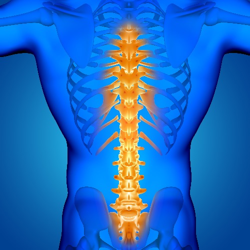
Spina bifida, known as the neural tube defect, is a congenital condition where the spine does not develop properly, leaving a gap in the spinal cord or surrounding vertebrae. The neural tube is a critical structure in embryonic development, forming early in pregnancy and eventually developing into the baby’s brain, spinal cord, and the protective tissues surrounding them. When the neural tube fails to close properly during the first few weeks of gestation, it can result in spina bifida. This condition can range from mild, with no visible symptoms, to severe forms that cause significant neurological and physical challenges. Surgery is often necessary to close this gap, protect the spinal cord, and prevent further complications.
Spina bifida occurs in three main forms: spina bifida occulta (mild), meningocele, and myelomeningocele(severe). Spina Bifida Occulta is the mildest form, involving a small gap in the vertebrae. Often asymptomatic, it’s commonly discovered through imaging tests like X-rays. Myelomeningocele, also known as “open spina bifida”, is the most severe type, with the spinal cord and nerves protruding through an open vertebra, forming a sac on the baby’s back. This exposes nerves and tissues. Meningocele is a rare type where a sac of spinal fluid protrudes through the spine without involving nerves.
Fetal surgery and postnatal surgery are the two main spina bifida treatment options. Choosing between prenatal or postnatal spina bifida repair involves evaluating gestational age, the location and severity of the myelomeningocele lesion, and maternal health consideration.


